DraftNot yet reviewed by the clinical team · Last edited 14 October 2026
Nephrostomy tube
When urine cannot drain out of a kidney, a nephrostomy gives it another way out: a thin, soft tube through the skin of your back into the kidney. It is usually done while you are awake, with numbing medicine and medicine to help you relax, and it eases the pressure right away.
The short version
- Something is blocking the tube that carries urine from your kidney to your bladder, so urine is backing up and the kidney is swelling.
- A nephrostomy is a thin, soft tube that goes through the skin of your back into the kidney, so the urine can drain into a bag outside your body.
- You lie face down and usually stay awake, with numbing medicine and medicine to help you relax. The doctor uses ultrasound and X-ray to guide a needle, a wire and then the tube.
- The tube eases the pressure right away and stays in until the blockage is fixed, and the team will teach you how to care for it at home.
What it treats
Your kidneys make urine all day
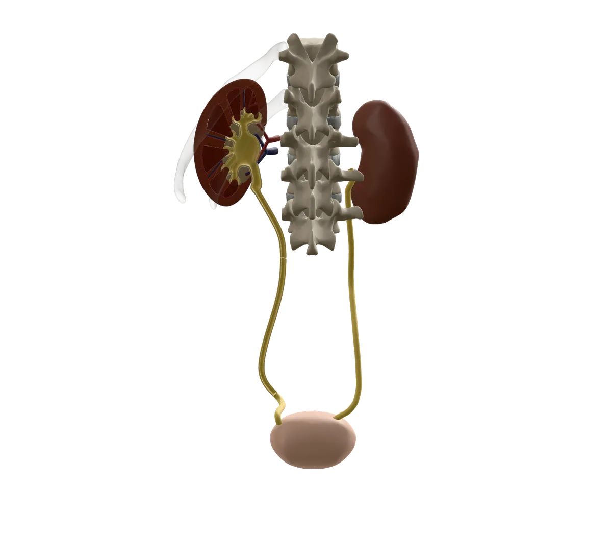
Your kidneys sit at the back of your belly, just below your ribs, one on each side of your spine. Each one is about the size of a fist. They clean your blood and turn the waste into urine (pee).
Urine collects in a small funnel in the middle of each kidney. From there it runs down a thin tube, called the ureter, to your bladder.
A blockage makes the kidney swell
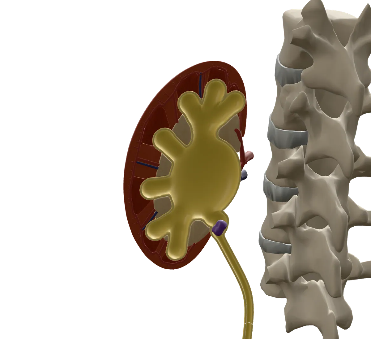
Sometimes something blocks the ureter. It might be a kidney stone, a growth in or near the tube, or damage from an injury or past surgery. Urine backs up, and the kidney swells. Doctors call this hydronephrosis.
A swollen kidney can cause pain in your side or back. Trapped urine can also get infected, which can make you very sick. Over time, a blocked kidney can be damaged.
How it works
A tube gives the urine a way out
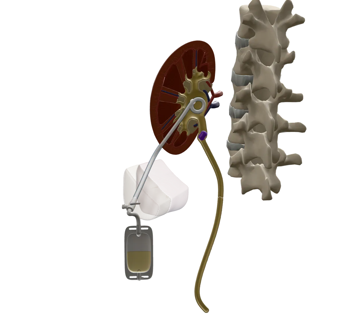
A nephrostomy is a thin, soft tube that goes through the skin of your back into the kidney. Urine drains out through the tube into a bag outside your body.
This takes the pressure off the kidney right away. It does not remove the blockage. It buys time, so your kidney can recover while your team plans how to fix the cause.
What the model shows
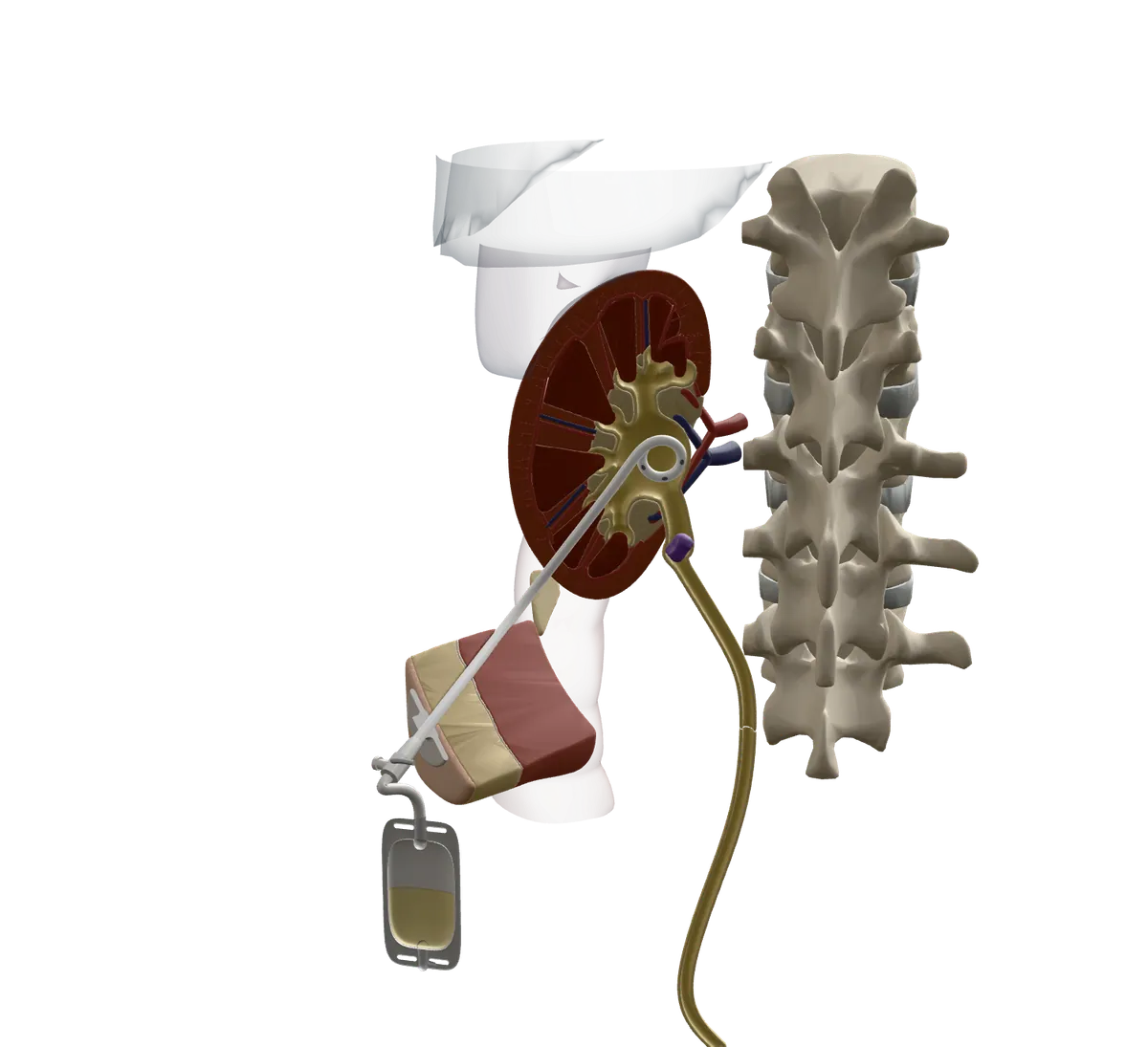
The model is a block of the left lower back, seen from behind, the way the doctor sees it when you lie face down. The kidney is cut open so you can see inside it.
The bowel, spleen and lung lining are see-through. They are there because they sit close to where the tube goes in.
The procedure
Getting ready
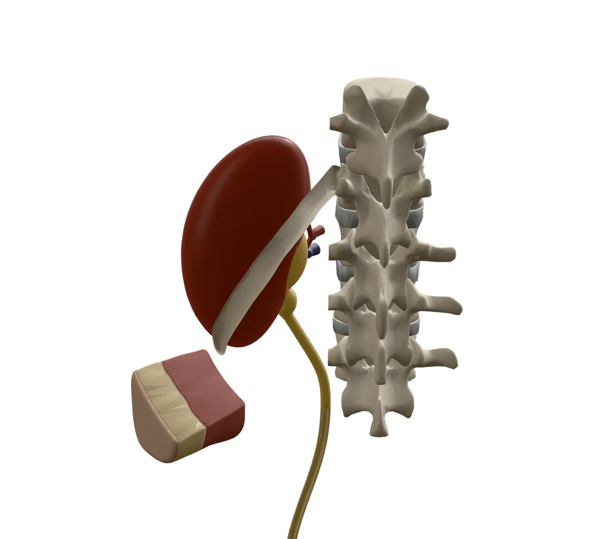
You will have blood tests to check that your blood clots well.
Often this is done quickly, because the kidney is infected or not working well. A nurse puts a small IV line in your arm. You get antibiotics through it to help prevent infection.
Good to know. Tell the team about all your medicines, especially blood thinners, and about allergies, especially to X-ray dye. Your team will tell you when to stop eating and drinking.
You lie face down, awake and numb
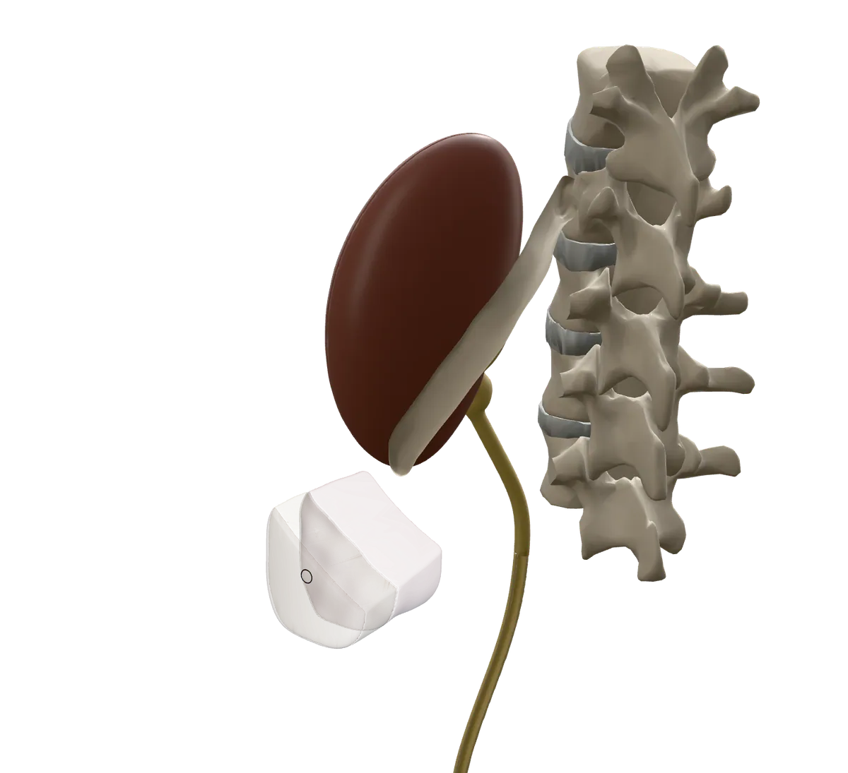
You lie on your stomach on the X-ray table. Most people get medicine through the IV to feel relaxed and drowsy. The doctor numbs the skin and the deeper tissue of your back.
The doctor uses ultrasound and X-ray to find the kidney and plan the path.
Good to know. You may feel some pressure, pushing or discomfort. Tell the team if it hurts.
A thin needle goes into the kidney
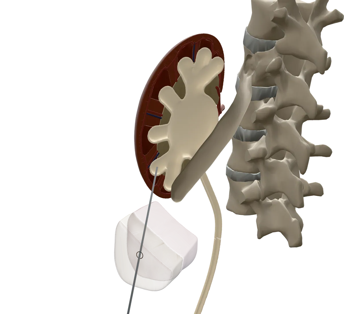
The doctor guides a thin needle through your back into one of the small cups inside the kidney. The best spot is usually a cup at the back of the middle or lower part of the kidney. The needle goes in below your lowest rib.
This path stays away from the larger blood vessels and from the lining of the lung. A little X-ray dye goes in through the needle, so the inside of the kidney shows on the screen.
A wire guides the soft tube into place
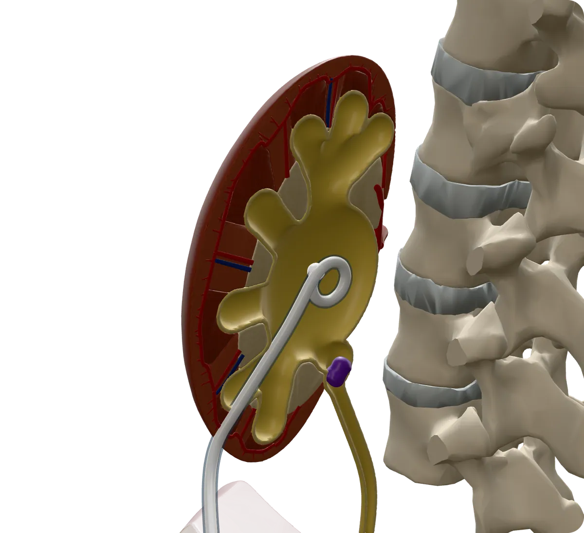
A thin, bendy wire goes through the needle into the kidney, and the needle comes out. Then the soft tube slides in over the wire.
When the wire comes out, the end of the tube curls into a small loop inside the kidney. The loop helps keep the tube from slipping out.
The tube is fixed to your skin and joined to a bag
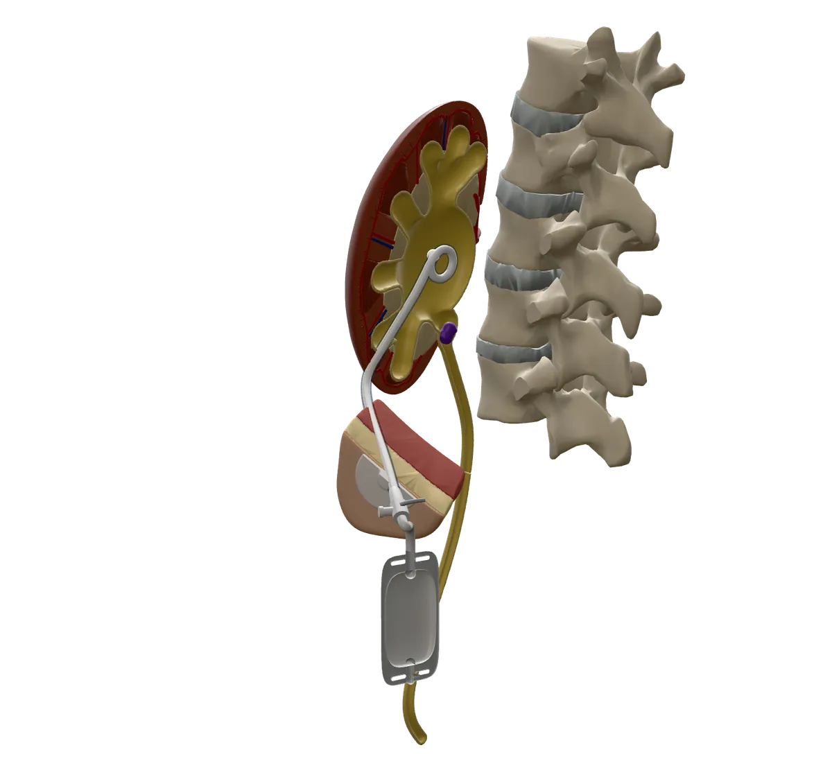
More dye checks that the tube is in the right place. The tube is held to your skin with a stitch or a sticky pad, covered with a dressing, and joined to a drainage bag. There is no surgical cut.
Good to know. The procedure usually takes about 20 minutes to an hour. Afterwards you rest in bed for a few hours. Some people go home the same day. Many stay in hospital for the illness that caused the blockage.
Afterwards
The kidney can drain again
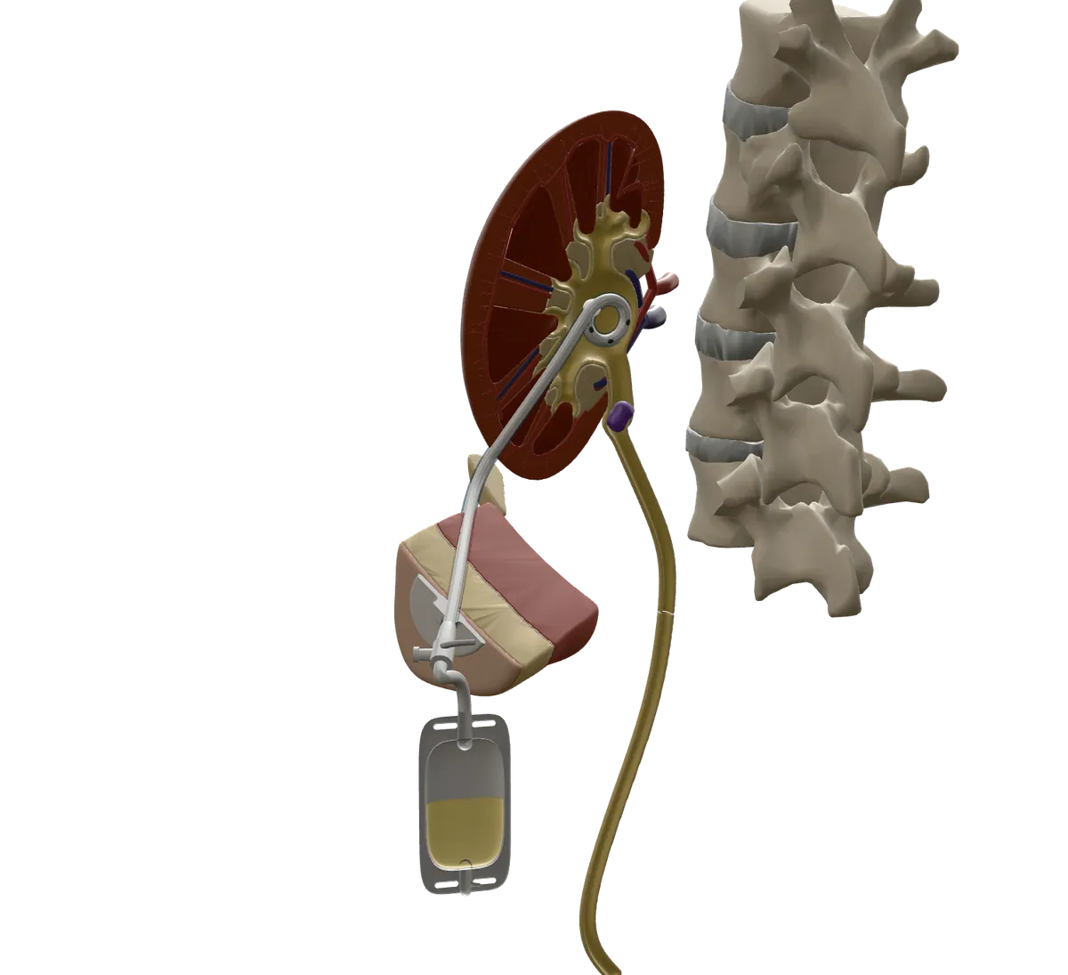
Urine now flows out through the tube. The pressure eases, and the swelling goes down. If you had an infection, your fever and pain should start to settle.
Pink urine in the bag is common for the first day or two.
Good to know. Your back may be sore for a week or two. If only one kidney has a tube, you will usually still pee the normal way too.
Living with the tube and bag
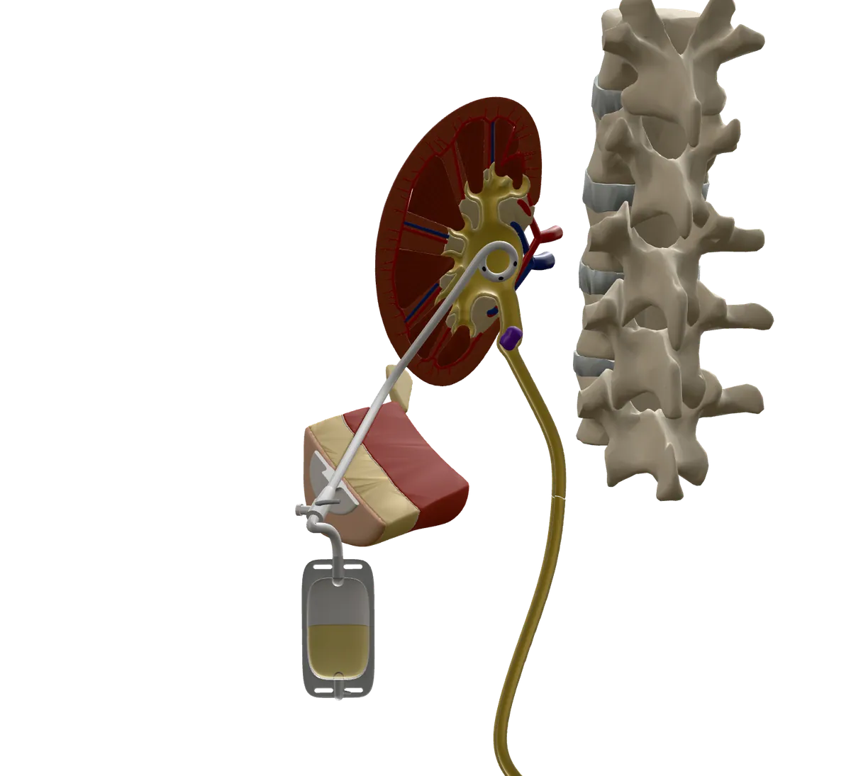
Keep the bag below your kidney, and empty it when it is half to two-thirds full. Keep the skin clean and dry, and change the dressing when your team says, or if it gets wet or loose.
Most people can shower after about 2 days, with the spot covered in plastic wrap. Do not swim or soak in a bath.
Good to know. Use a fresh bag about once a week. Only flush the tube if your team showed you how. If you need the tube for a long time, it is changed about every 2 to 4 months.
Risks
Risks, explained plainly
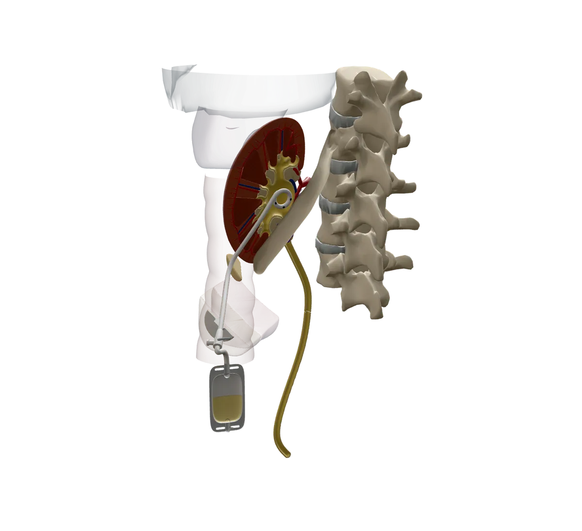
Every procedure has some risk, and most people do not have a serious problem. Each risk is listed under Benefits and risks below, with how often it happens.
Most people have no serious problems, but about 1 to 4 in 100 people have bleeding that needs a blood transfusion.
Your visit
Before, on the day and after. Your care team’s instructions come first if they differ.
Before your procedure
Things to do. Tick them off as you go; your ticks stay on this device only.
On the day
You change into a gown, and a nurse puts a small IV line in your arm.
You get antibiotics through the IV to help prevent infection.
You lie face down, or on your side, on the X-ray table.
Medicine through the IV helps you relax. The doctor numbs the skin and deeper tissue of your back.
Using ultrasound and X-ray, the doctor places a thin needle, then a wire, then the tube.
The tube is held with a stitch or a sticky pad and joined to a bag. This takes about 20 minutes to an hour.
You rest in bed for a few hours. Some people go home the same day, with someone to drive them. Many stay in hospital.
Before you leave, a nurse shows you how to empty the bag and care for the tube.
Recovery
- The first few hours
Rest in bed while nurses check you and the tube. Pink urine in the bag is normal. Tell the nurse before you get up, and move slowly so the tube does not get pulled.
- Days 1 and 2
Blood in the urine usually clears within a day or two. A little clear or light yellow fluid around the tube is normal for 1 to 3 days. Take it easy, and avoid sudden moves.
- The first 1 to 2 weeks
Almost everyone has some soreness in their back where the tube goes in. This is expected and usually eases over 1 to 2 weeks. Acetaminophen may help. Ask before taking ibuprofen or aspirin, because they can add to bleeding. Most people are back to light, everyday activity within a few days.
- Every day with the tube
Keep the bag below your kidney, empty it at half to two-thirds full, check for kinks, and keep the skin clean and dry. Drink at least 2 litres of fluid a day unless your team says otherwise.
- Every 2 to 4 months
If you need the tube for a long time, it is changed for a new one, usually as an outpatient visit. If you have not heard about a change by 3 months, call the team.
- When the problem is fixed
The tube may be closed for about a day to test the kidney, then taken out after numbing. Some people get an internal stent instead, with no bag. Avoid hard exercise, baths and swimming for a few days after the tube comes out.
When to get help
Call 9-1-1 now if you have
- Breathing very fast.
- Confusion, slurred speech, or not making sense.
- Blue, pale or blotchy skin.
- A very high or very low temperature, feeling hot or cold to the touch, or shivering.
- A rash that does not fade when you press it.
- Heavy bleeding from the tube or from the skin around it.
- Anything else that feels like an emergency.
Call the interventional radiology (IR) team today if you have
- The tube falls out or slips. Cover the hole with a clean dressing first.
- No urine comes into the bag.
- Fever or chills.
- Bright red urine, like beet juice, or blood in the urine after the first few days.
- New or worse pain in your back or side.
- Urine leaking around the tube.
- Redness, swelling, pus or a bad smell where the tube goes in.
- Cloudy or bad-smelling urine.
- Throwing up.
- The stitch or pad is pulling away from your skin.
Phone number added by the hospital before launch
If you can’t reach them, go to the nearest emergency department.
Not sure? Call HealthLink BC at 8-1-1
- You are not sure how urgent something is.
- You cannot reach the IR team.
- You have a question that is not urgent, like mild skin irritation under the tape.
A nurse can give health advice any time, day or night.
Call 8-1-1Deaf or hard of hearing: call 7-1-1. Translation services are available in over 130 languages.
Benefits and risks
What it can do for you
- It eases the pressure on your kidney right away, so the kidney can start to recover.
- In a swollen, blocked kidney, the tube is placed successfully 96 to 100 times out of 100.
- If the kidney is infected, draining it is a key step in treating the infection.
- For severe infection from a kidney stone, it works about as well as an internal stent.
- No surgical cut, and usually no general anesthetic (medicine that puts you to sleep).
- The same opening can later be used to place an internal stent, so you may not need a bag for long.
What can go wrong
Every procedure has some risk, and most people do not have a serious problem. Each risk is listed with how often it happens. They are grouped on one scale, from very common to very rare.
Very common: more than 1 in 10 people
Infection and sepsis
A UK patient leaflet says any spreading infection happens to between 1 in 10 and 1 in 2 people. The most serious form (septic shock, when blood pressure drops) happens to about 1 to 10 in 100 people. Septic shock happens to 7 to 9 in 100 if the kidney was already full of pus.
If the trapped urine was infected, germs can get into the blood when the kidney is drained. This is called sepsis, and it is serious. You get antibiotics first to lower the risk. The risk is higher if the kidney was already full of pus. Sources count different things, so the numbers differ.
Tube blockage
Between 1 in 10 and 1 in 2 people.
Crust, blood clots or bits of tissue can block the tube, so urine stops draining. It may need flushing or a new tube.
Leaking or slipping out
Leaking happens to between 1 in 10 and 1 in 2 people. Slipping out happens to fewer than 1 in 100 people in the first days. Over the following months, it happens to 11 to 30 in 100.
Urine can leak around the tube where it leaves the skin. The tube can also be pulled or slip out, and would need to be put back.
Common: 1 in 10 to 1 in 100 people
Bleeding
Bleeding that needs a blood transfusion happens to about 1 to 4 in 100 people. Bleeding that needs a procedure to seal an artery happens to about 1 to 10 in 1,000.
The tube passes through kidney tissue, which has many small blood vessels. Pink urine for a day or two is normal. A few people bleed more, and need a blood transfusion or a procedure to seal a bleeding artery.
Nearby organs
A UK patient leaflet says between 1 in 250 and 1 in 50 people have an injury to a nearby organ, including the lung. The bowel is hurt in about 2 to 5 in 1,000 people.
Once in a while, the needle goes through the bowel, which sometimes lies beside or behind the kidney. The spleen and liver sit near the top of the kidneys. Only a few cases of injury to them have been reported.
Uncommon: 1 in 100 to 1 in 1,000 people
Lung lining
About 1 to 6 in 1,000 people.
The bottom edge of the lung lining sits behind the lowest ribs. If the needle crosses it, air or fluid can collect around the lung. Going in below the lowest rib keeps this uncommon.
Living with the tube
Other options
Questions to ask
Tick the ones you want to ask. Your ticks stay on this device only.
Check your understanding
Nothing is saved. It is just for you.
It gives trapped urine a way out, so the pressure eases. The blockage itself is treated separately.
Below the kidney, so urine flows down into the bag and does not back up.
The team may need to put in a new tube. If you cannot reach them, go to the emergency department.
These can be signs of sepsis, a serious infection. Pink urine on the first day is common.
Cover the spot with plastic wrap and change a wet dressing. Do not swim or soak in a bath while the tube is in.
Words used
- Kidney
- One of two bean-shaped organs at the back of the belly that clean the blood and make urine.
- Ureter
- The thin tube that carries urine from each kidney down to the bladder.
- Renal pelvis
- The funnel in the middle of the kidney where urine collects before it goes down the ureter.
- Calyx
- One of the small cups inside the kidney that catch urine and empty into the renal pelvis. More than one are called calyces.
- Hydronephrosis
- A kidney that is swollen because urine cannot drain out.
- Nephrostomy
- A tube placed through the skin into the kidney to drain urine into a bag.
- Percutaneous
- Done through the skin with a needle, not a surgical cut.
- Catheter
- A thin, flexible tube. Your nephrostomy tube is a kind of catheter.
- Guidewire
- A thin, bendy wire the doctor uses to guide the tube into place.
- Ureteric stent
- A thin tube placed inside the ureter so urine can drain to the bladder with no bag.
- Sepsis
- A serious reaction to infection that affects the whole body. It is an emergency.
- Interventional radiologist
- A doctor who treats problems through tiny openings, guided by X-ray, ultrasound or CT.
Sources
- Society of Interventional Radiology. Quality Improvement Guidelines for Percutaneous Nephrostomy (J Vasc Interv Radiol, 2016)
- Seminars in Interventional Radiology. Percutaneous Nephrostomy: Technical Aspects and Indications (2011)
- Cardiovascular and Interventional Radiological Society of Europe (CIRSE). Nephrostomy and ureteric stenting, patient information
- British Society of Interventional Radiology. Percutaneous Nephrostomy, patient information
- British Association of Urological Surgeons. Percutaneous insertion of a nephrostomy tube, patient leaflet (2023)
- RadiologyInfo.org (RSNA and ACR). Ureteral Stenting and Nephrostomy
- Canadian Association for Interventional Radiology. Nephrostomy drainage
- Canadian Association for Interventional Radiology. Nephrostomy catheter care
- Cleveland Clinic. Nephrostomy Tube: Care, Purpose and Complications
- Cleveland Clinic. Hydronephrosis
- Cleveland Clinic. Obstructive Uropathy
- MedlinePlus, US National Library of Medicine. Percutaneous urinary procedures, discharge (2024)
- NHS hospital trust patient leaflet (England). Caring for your Nephrostomy Tube at Home (version 6, 2024)
- NHS hospital trust patient leaflet (England). Nephrostomy tube, patient information (2022)
- British Association of Urological Surgeons. Nephrostomy troubleshooting (clinician guide)
- Medicina (peer-reviewed journal). Percutaneous Nephrostomy versus Ureteral Stent for Severe Urinary Tract Infection with Obstructive Urolithiasis: A Systematic Review and Meta-Analysis (2024)
- Seminars in Interventional Radiology. Nephroureteral Stents: Principles and Techniques (2011)
- Indian Journal of Nephrology. Percutaneous nephrostomy by direct puncture technique: An observational study (2010)
- National Institute of Diabetes and Digestive and Kidney Diseases. Your Kidneys and How They Work
- NHS. Sepsis
- HealthLink BC. 8-1-1 health advice and emergencies
- NHS. Hydronephrosis (reviewed July 2025)
- MedlinePlus, US National Library of Medicine. Obstructive uropathy
- Macmillan Cancer Support. Nephrostomy
- HealthLink BC. When to visit the emergency room (updated January 2025)
- HealthLink BC. HealthLink BC 8-1-1 services
About this guide
- Status
- Draft: not yet reviewed by the clinical team. The interventional radiology team will review it before it is used with patients, and the date of that review will show here.
- How it was written
- Drafted with AI writing tools from 25 published sources, then fact-checked against those sources by a separate AI check. Anything the sources did not support was taken out. The interventional radiology team will review it before it is used with patients.
- Last edited
- 14 October 2026
- Document
- Pathway P-005 · Version 0.5 · Last reviewed: not yet · Next review: 12 months after sign-off · Owner: the interventional radiology team
Was this guide clear?
Tell us if anything was hard to follow, or if something on the page looks wrong. Please do not include details about your own health. For questions about your care, contact your care team.
Both open an email to the project team. Project email: added before launch