DraftNot yet reviewed by the clinical team · Last edited 14 October 2026
Endovascular aneurysm repair
EVAR repairs a bulge in the body’s main artery from the inside, through small openings at both groins. A fabric tube on a thin metal frame takes the pressure off the weak wall, with no cut in the belly.
The short version
- An abdominal aortic aneurysm is a weak, bulging part of the body’s main artery, and a large one can burst.
- In EVAR, the team reaches the aorta through both groins and slides a fabric and metal tube inside it, guided by live X-rays.
- The tube opens across the bulge, so blood flows through the tube and stops pushing on the weak wall.
- Most people go home in 1 to 3 days, but you need scans for life to check that the tube stays sealed.
What it treats
The aorta is your body’s main artery
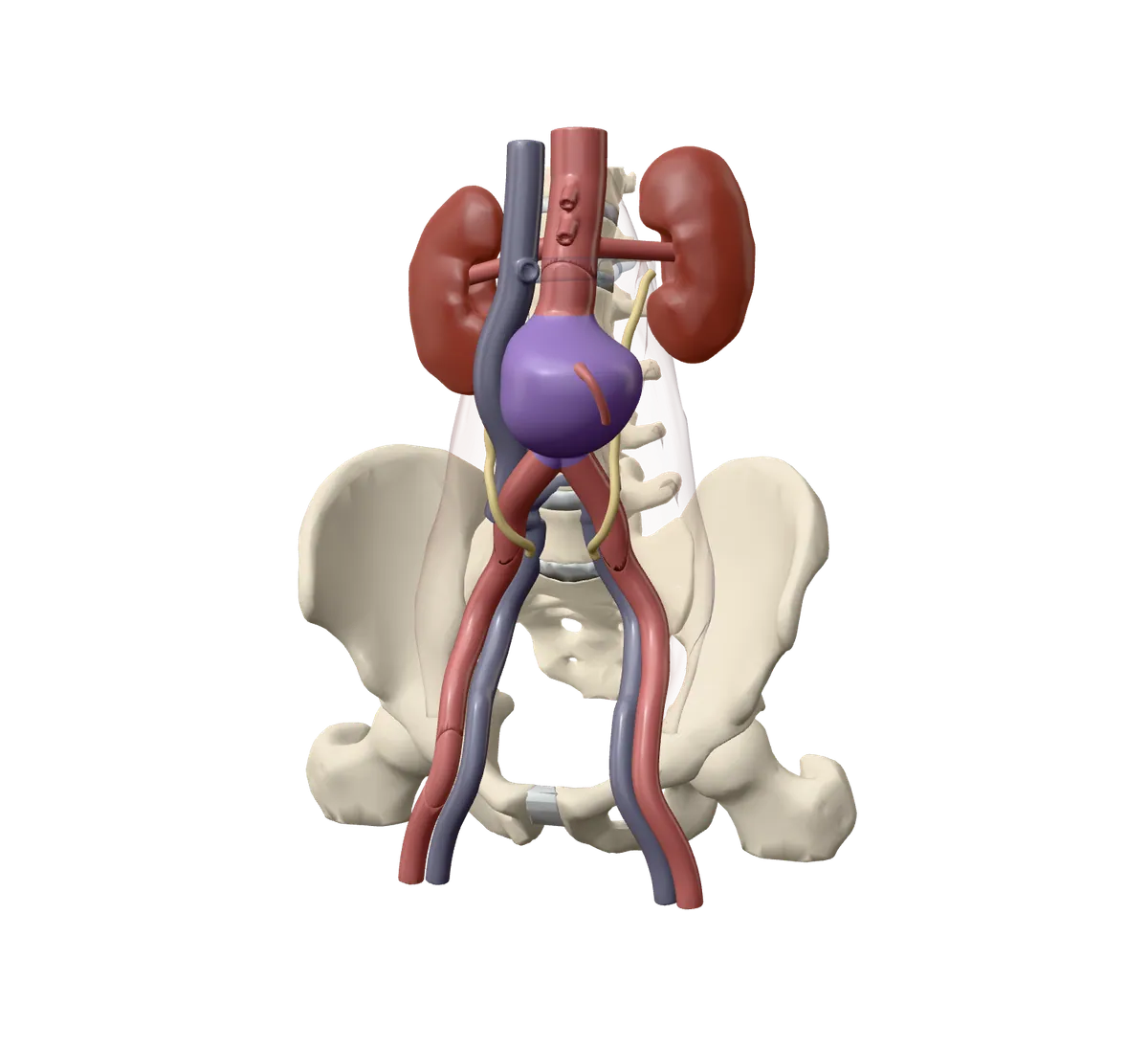
An artery is a blood vessel that carries blood away from the heart. The aorta is the biggest one. It runs from your heart down through your belly, then splits in two to feed your legs.
In most adults, the aorta in the belly is about 2 cm wide. It lies in front of the spine, between the kidneys, and splits in two low in the belly.
An aneurysm is a weak spot that bulges
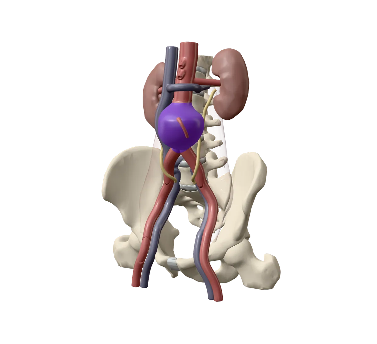
An aneurysm (say AN-yur-iz-um) is a part of an artery wall that has become weak and stretched, like a balloon. In the belly, it is called an abdominal aortic aneurysm, or AAA.
Most cause no symptoms and are found by chance on a scan.
Good to know. They are more common in men over 65 and in people who smoke or used to smoke. Having a close relative who had one also raises the chance.
A large bulge can burst, so it is fixed first
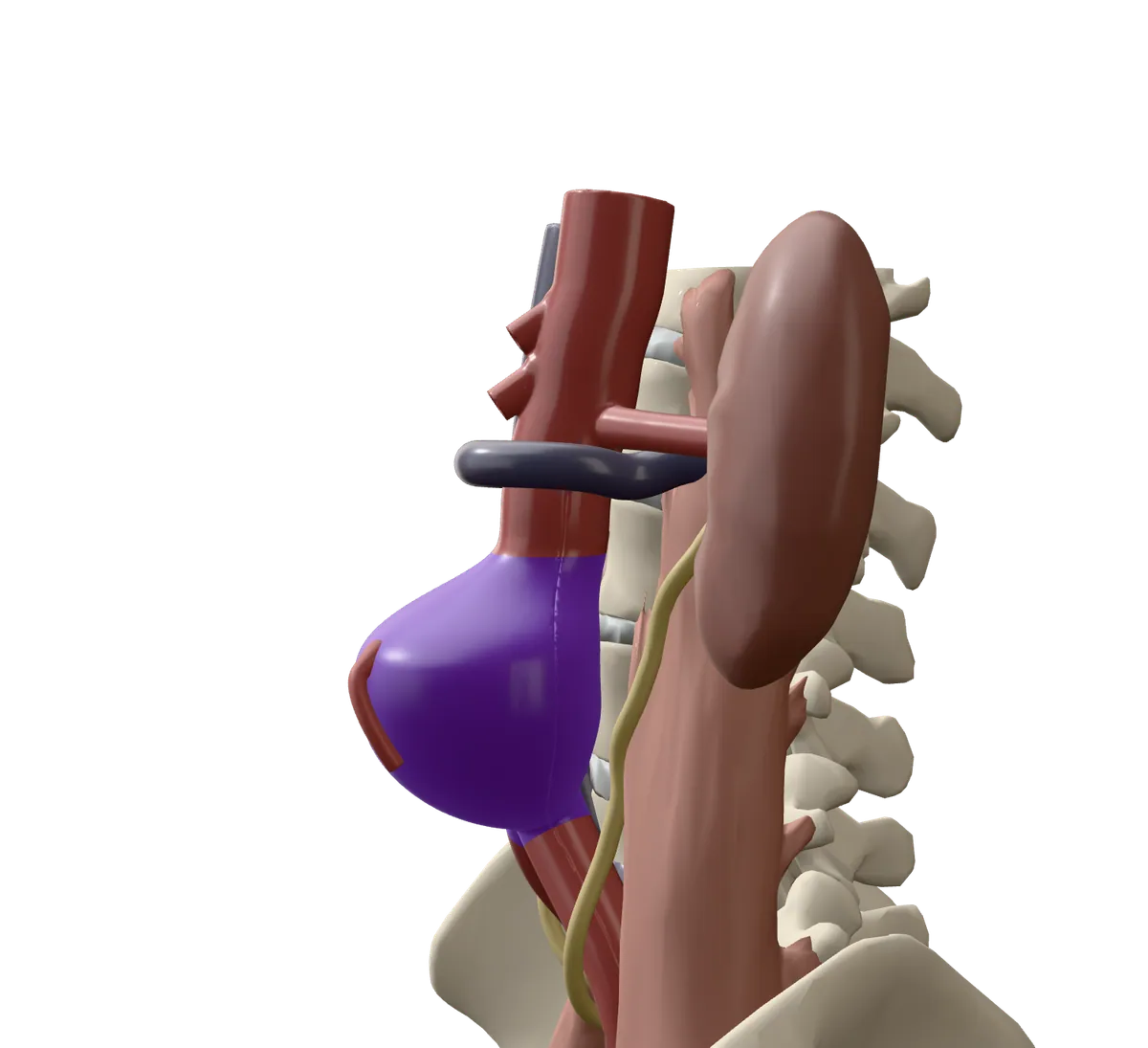
A bulge that keeps growing can burst. This is called a rupture. It causes severe bleeding inside the body, and most people do not survive it.
Small aneurysms rarely burst, so they are usually watched with scans. Repair is often offered when the bulge reaches about 5.5 cm, grows quickly, or causes pain. Your team weighs the risk from the bulge against the risk of the repair.
How it works
A stent graft lines the artery from the inside
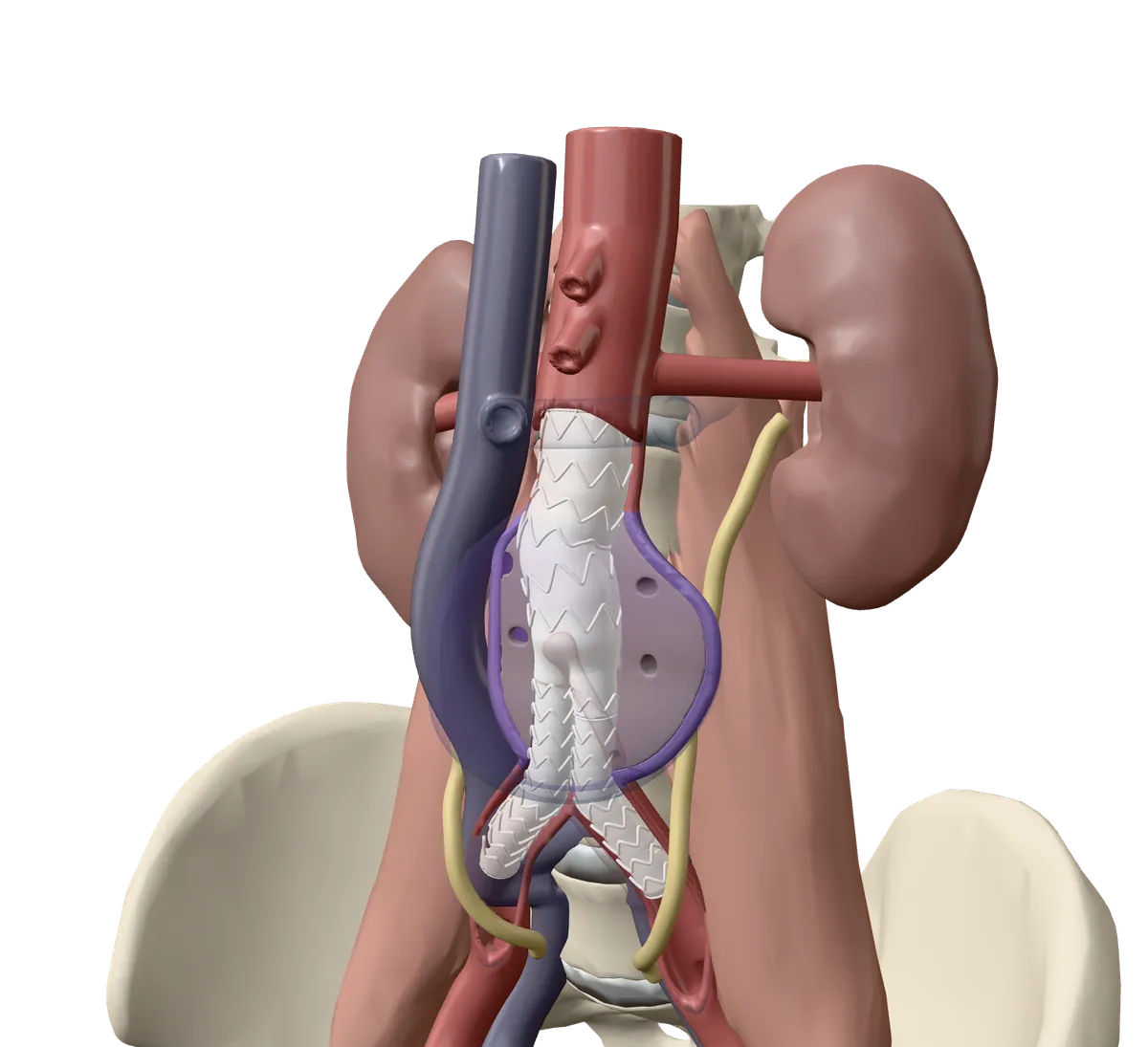
In EVAR, the bulge is not cut out. Instead, a tube called a stent graft is placed inside the aorta. It is made of strong fabric held open by a thin metal frame.
Blood flows through the new tube instead of pushing on the weak wall. The tube splits in two near the bottom, like a pair of trousers, with one leg going into each leg artery.
The graft comes in pieces that join inside you
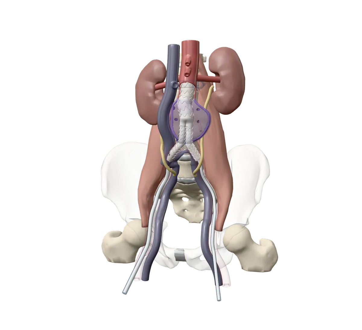
The graft goes in folded up small inside a thin tube. Once it is in the right place, it opens and presses against the healthy artery wall above and below the bulge. Small hooks help hold the top in place.
The pieces are put together inside the body. The main body goes in first from one groin. Then a second leg goes in from the other groin and joins onto it.
The procedure
Getting ready: scans, tests and planning
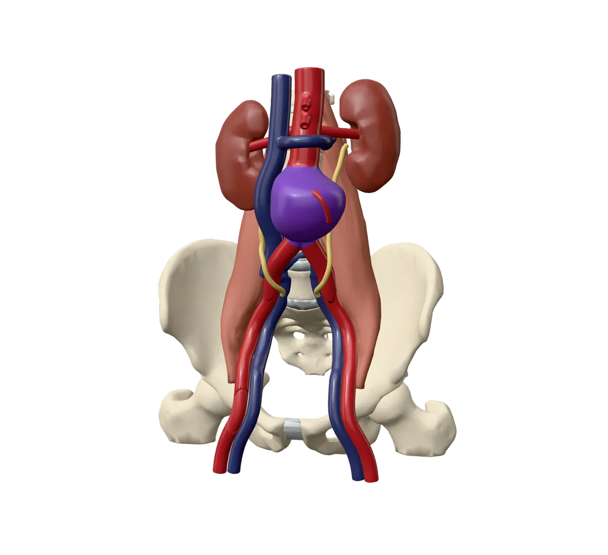
A CT scan, a detailed X-ray scan, measures your aorta so the team can choose a graft that fits. You will likely have blood tests and a heart tracing (ECG). If you have ever reacted to X-ray dye, tell the team.
Good to know. Bring a list of your medicines. Your team will tell you which ones to stop or change. You will be told when to stop eating before the procedure. Plan for someone to drive you home.
The way in is through both groins
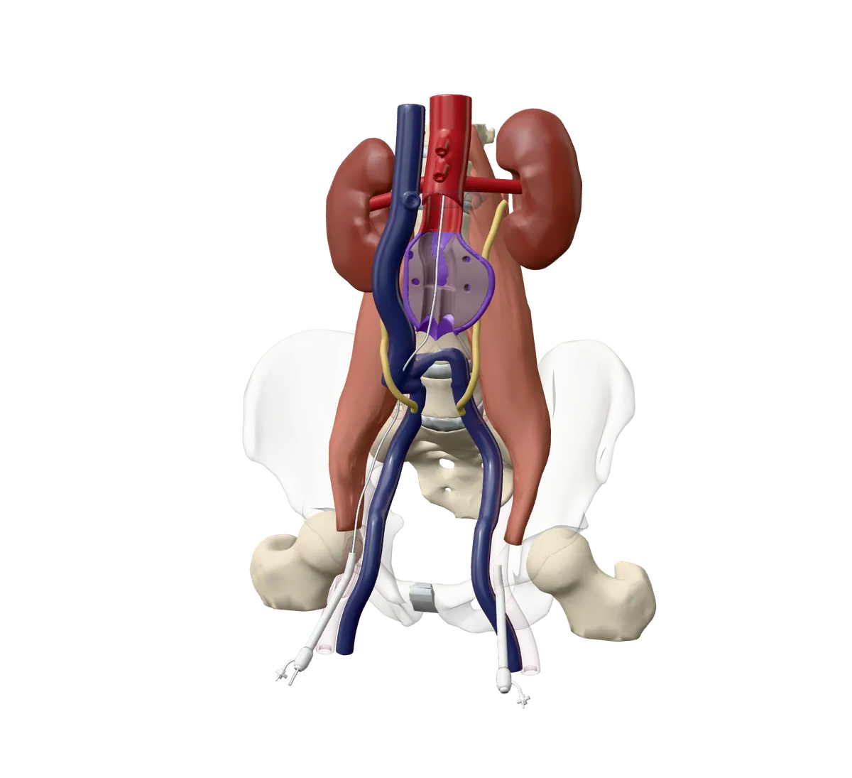
You will be asleep, or awake and numbed (from the waist down or just at the groins) with medicine to help you relax.
The artery at each groin is reached with a needle opening or a small cut. A short tube called a sheath goes into each artery, and a thin, soft wire is passed up to the aorta. You will not feel the wire inside the artery.
Good to know. The team works in an operating room or an X-ray room with live X-ray screens. An interventional radiologist and a vascular surgeon often work together.
The main body opens just below the kidney arteries
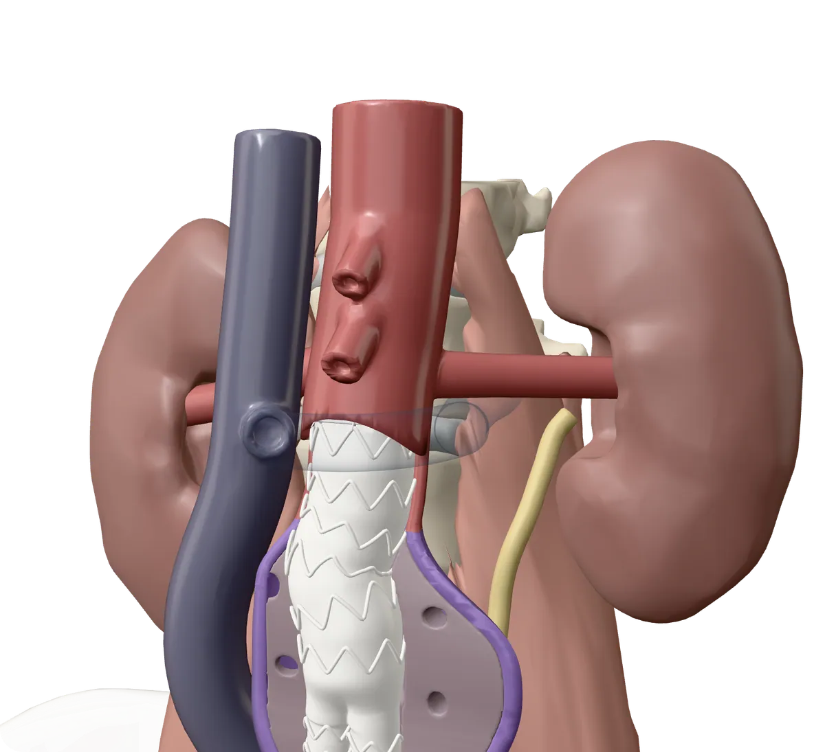
The team injects X-ray dye to map the aorta and the kidney arteries. You may feel a warm flush for a moment.
The folded main body slides up over the wire. When it sits just below the kidney arteries, the team opens it. It springs out and seals against the healthy artery wall.
Good to know. If you are awake, you may be asked to hold your breath for short periods.
The second leg joins, and the seal is checked
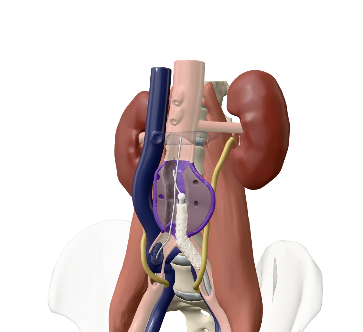
From the other groin, the team steers a wire into a short opening on the main body. The second leg slides in and opens there, so the two pieces overlap.
A last X-ray with dye checks that blood flows only through the graft and not into the bulge.
The tubes come out and the groins are closed
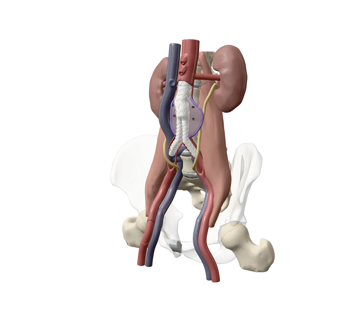
The wires and sheaths are removed. Each artery is closed with stitches or a small closing device, and the skin is closed or covered.
You go to a recovery area to wake up and be watched closely.
Good to know. The whole procedure usually takes about 2 to 3 hours. Most people stay in hospital for 1 to 3 days.
Afterwards
The bulge is checked for the rest of your life
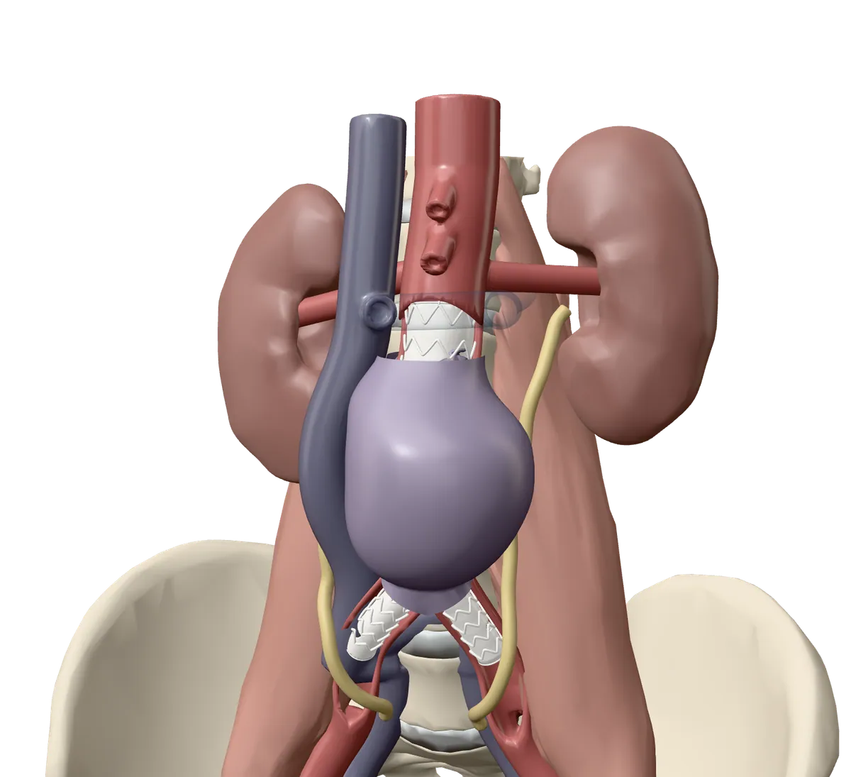
The bulge stays in your body, but blood no longer pushes on it. In about half of people, it is smaller after a year. In others it stays about the same size, and your team keeps watching it.
You will need scans, often a CT scan or an ultrasound, for the rest of your life. They check that the graft has not moved and that no blood is leaking into the bulge.
Risks
Risks, explained plainly
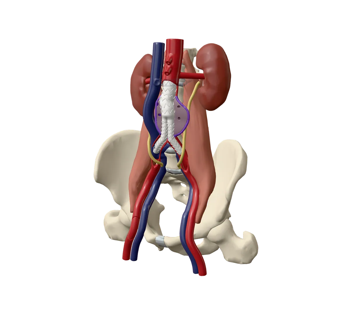
Every procedure has some risk, and most people do not have a serious problem. Each risk is listed under Benefits and risks below, with how often it happens.
Fewer than 2 in 100 people die around the time of EVAR, and about 1 to 2 in 10 need another, usually smaller, procedure later.
Your visit
Before, on the day and after. Your care team’s instructions come first if they differ.
Before your procedure
Things to do. Tick them off as you go; your ticks stay on this device only.
On the day
Arriving. You may come in on the day, or the day before. You change into a gown, and a small IV line (a thin tube) is put in a vein in your arm.
Consent. The team explains the procedure again and asks you to sign a consent form. You can still ask questions, or decide not to go ahead.
Anesthetic. You will be asleep (general anesthetic), or numbed from the waist down or at the groins, with medicine to help you relax.
The room. An X-ray room or operating room with large screens. An interventional radiologist and a vascular surgeon often work together.
How long. About 2 to 3 hours, sometimes longer.
Waking up. You wake up in a recovery area. You may have a thin tube draining your bladder until the next day.
Recovery
- The first hours
Nurses check your blood pressure, pulse and groins often. You may need to keep your legs straight for a few hours. You can usually eat and drink soon after.
- Days 1 to 3
Most people walk the same evening or the next day, and go home in 1 to 3 days. The groins are sore and bruised. A short spell of fever and tiredness can happen.
- Weeks 1 to 2
Groin soreness and tiredness ease. Many daily activities can start again. Walk a little more each day.
- Weeks 2 to 6
Full recovery can take up to 4 to 6 weeks. Avoid heavy lifting until your team says it is okay (advice ranges from 2 to 6 weeks). Drive only when you can do an emergency stop without pain and are off pain medicine.
- Around 6 weeks
In some hospitals, the first check-up and scan are around this time.
- For life
Regular scans, at least once a year, to check the graft. Take aspirin and a cholesterol medicine if they are prescribed. Tell your dentist and other doctors that you have a graft in your aorta.
When to get help
Call 9-1-1 now if you have
- Sudden, severe pain in your belly, back or groin
- Fainting, collapsing, or feeling very dizzy
- Chest pain
- Bad trouble breathing
- A lump at the groin that is quickly getting bigger
Call the interventional radiology (IR) team today if you have
- A groin wound that is red, hot, swollen, oozing, or more painful each day
- Bright red blood soaking through the dressing
- A fever over 37.8°C
- Pain that does not get better with your pain medicine
- A cold foot, or a toe that turns blue or black
- Swelling, pain or redness in a leg (this can be a blood clot)
- New back or belly pain that is not severe
Phone number added by the hospital before launch
If you can’t reach them, go to the nearest emergency department.
Not sure? Call HealthLink BC at 8-1-1
- You are not sure if something is normal
- Questions about wound care, medicines or activity when your team is closed
- Non-urgent advice from a nurse, any time of day
A nurse can give health advice any time, day or night.
Call 8-1-1Deaf or hard of hearing: call 7-1-1. Translation services are available in over 130 languages.
Benefits and risks
What it can do for you
- No cut in the belly. The graft goes in through two small openings, one at each groin.
- Less risk of dying around the time of the repair than with open surgery: fewer than 2 in 100 people, compared with about 4 in 100, when four large trials were combined.
- A shorter hospital stay: most people go home in 1 to 3 days. In one large trial, the middle stay was 3 days, compared with 7 days after open surgery.
- Less blood loss and a shorter procedure than open surgery.
- A faster recovery: many daily activities can start again after 1 to 2 weeks.
- It works: the bulge is kept from bursting in more than 99 in 100 people, according to one specialist society.
- To be fair: after a few years, people live about as long after EVAR as after open surgery, and more people need a second procedure after EVAR.
What can go wrong
Every procedure has some risk, and most people do not have a serious problem. Each risk is listed with how often it happens. They are grouped on one scale, from very common to very rare.
Very common: more than 1 in 10 people
A leak from a small side artery (type II endoleak)
It is seen in about 10 to 20 in 100 people at the 1-month scan. About 5 to 15 in 100 still have one after 6 months. Between 30 and 75 in 100 of these leaks seal on their own.
Small arteries that used to feed the bulge can let blood seep back into it, around the outside of the graft. Many seal on their own, so the team watches them on your scans. They are treated if the bulge grows.
Needing another procedure later
About 15 in 100 people within 4 years.
Where this number comes from
About 15 in 100 people within 4 years, says one patient leaflet. Other sources say about 1 in 10.
Because leaks or movement can happen months or years later, some people need a second, usually smaller, procedure.
A flu-like spell
About 3 in 10 people.
Where this number comes from
About 3 in 10 people, in combined studies of stent grafts in the chest and belly.
In the first days, some people have a fever, aches and tiredness as the body reacts to the graft. It is usually not serious, but it can mean a longer stay in hospital.
Common: 1 in 10 to 1 in 100 people
Bleeding, bruising or infection at the groin
About 5 in 100 people had a problem at the groin opening.
Where this number comes from
About 5 in 100 people had a problem at the groin opening, in a large registry of about 34,000 people.
Bruising and soreness at the groin are common for a week or two. Sometimes a larger lump of blood under the skin (a haematoma), a clot, or a wound infection happens. In severe cases, another small operation is needed. Problems at the groin are more common in women (about 8 in 100) than in men (about 4 in 100).
A leak at a seal, or the graft moving (type I endoleak)
About 8 in 100 people had a leak at the top seal, in one study of 389 people followed for about 7 years. On average it showed up about 4 years after the repair.
If the top or bottom of the graft does not seal tightly, or it slips over time, blood can get into the bulge at full pressure. This is more serious and needs to be fixed quickly, often with another catheter procedure. It is more likely when the healthy part of the aorta below the kidney arteries is wide or bent.
Strain on the kidneys
About 3 to 9 in 100 people, in two studies of standard EVAR. Fewer than 1 in 100 needed dialysis (a machine that cleans the blood), in one recent registry.
The X-ray dye can strain the kidneys, especially if they are already weak. It often gets better, but for some people the kidneys stay weaker afterwards. Your blood tests beforehand help the team plan for this.
A blocked graft leg
About 6 in 100 people, when 13 studies were combined. About half happen in the first month.
A clot can block one leg of the graft, so less blood reaches that leg. Most people need another procedure or an operation to fix it. More recent studies found it less often.
The bulge bursting after repair
6 of 444 people (about 1 in 75) had a burst within up to 9 years of EVAR.
Where this number comes from
6 of 444 people (about 1 in 75) had a burst within up to 9 years of EVAR, in one large trial.
Sometimes the bulge can still burst years later, usually because a leak or a moving graft was not found in time. This is why scans for life matter.
Serious problems and risk to life
About 1 to 2 in 100 people die around the time of EVAR, says HealthLink BC. One recent patient leaflet says fewer than 1 in 200. Graft infection happens to fewer than 1 in 100.
Rarely, a heart attack, stroke, chest infection, graft infection, or a problem with blood supply to the bowel, legs or spinal cord can happen. Any major procedure carries a small risk of death.
Other options
Questions to ask
Tick the ones you want to ask. Your ticks stay on this device only.
Check your understanding
Nothing is saved. It is just for you.
The bulge stays in place. A fabric and metal tube inside it takes the pressure off the weak wall.
The team uses a needle opening or a small cut at each groin. There is no cut in the belly.
Leaks or movement can happen months or years later. Scans find them early, when they are easier to fix.
Sudden, severe pain can be an emergency. Call 9-1-1 right away.
Moving and walking help you recover. Your team will tell you when heavy lifting is okay.
Words used
- Aorta
- The body’s main artery. It carries blood from the heart down through the chest and belly.
- Aneurysm
- A weak part of an artery wall that bulges out like a balloon.
- EVAR
- Endovascular aneurysm repair. Fixing an aneurysm from inside the blood vessel.
- Stent graft
- A fabric tube held open by a thin metal frame. It lines the artery from the inside.
- Endoleak
- Blood leaking into the bulge around the outside of the stent graft.
- Interventional radiologist
- A doctor who treats problems through tiny openings, guided by imaging such as X-rays.
- Vascular surgeon
- A surgeon who treats problems of the arteries and veins.
- Sheath
- A short, thin tube placed in the artery at the groin. Wires and the graft pass through it.
- Guidewire
- A very thin, bendy wire that the team steers through the arteries to lead the way.
- Contrast dye
- A liquid that shows up on X-rays, so the team can see the blood vessels.
- CT scan
- A detailed X-ray scan that shows the inside of the body in thin slices.
- Iliac arteries
- The two arteries the aorta splits into, one going toward each leg.
Sources
- HealthLink BC. Endovascular Repair for Abdominal Aortic Aneurysm
- HealthLink BC. Aortic Aneurysm
- Canadian Task Force on Preventive Health Care. Abdominal Aortic Aneurysm (2017)
- Cardiovascular and Interventional Radiological Society of Europe (CIRSE). Endovascular treatment of abdominal aortic aneurysms (EVAR), patient information
- Society of Interventional Radiology. Abdominal aortic aneurysm and IR treatment options (reviewed September 2024)
- RadiologyInfo.org (ACR and RSNA). Abdominal Aortic Aneurysm
- RadiologyInfo.org (ACR and RSNA). Catheter Angiography
- British Society of Interventional Radiology and The Royal College of Radiologists. Endovascular aortic repair (EVAR), patient leaflet (2011)
- NHS. Abdominal aortic aneurysm
- NHS. Abdominal aortic aneurysm screening
- Circulation Foundation (Vascular Society of Great Britain and Ireland). Endovascular aneurysm repair (EVAR)
- Circulation Foundation (Vascular Society of Great Britain and Ireland). Abdominal Aortic Aneurysm: Endovascular Aneurysm Repair (EVAR), patient leaflet
- NHS patient leaflet (Gloucestershire Hospitals NHS Foundation Trust). Endovascular Aneurysm Repair (EVAR), reviewed August 2025
- NHS patient leaflet (Oxford University Hospitals NHS Foundation Trust). Endovascular Aneurysm Repair (EVAR): information for patients, April 2026
- NHS patient leaflet (University Hospital Southampton NHS Foundation Trust). Endovascular aneurysm repair (EVAR), patient information factsheet, 2022
- Cleveland Clinic. Endovascular Aneurysm Repair (EVAR)
- NICE (NG156 decision support tool, published by NHS England). Making a decision about abdominal aortic aneurysm (AAA) (2023)
- JAMA (OVER trial). Outcomes following endovascular vs open repair of abdominal aortic aneurysm: a randomized trial (2009)
- New England Journal of Medicine (OVER trial). Long-term comparison of endovascular and open repair of abdominal aortic aneurysm (2012)
- The Lancet (EVAR trial 1). Endovascular versus open repair of abdominal aortic aneurysm in 15-years’ follow-up of the UK endovascular aneurysm repair trial 1 (2016)
- Cochrane Database of Systematic Reviews. Endovascular repair of abdominal aortic aneurysm (2014)
- Journal of the American Heart Association. Two decades of endovascular abdominal aortic aneurysm repair: enormous progress with serious lessons learned (2012)
- Journal of Clinical Medicine. Understanding type II endoleak: a harmless imaging finding or a silent threat? (2024)
- Journal of Vascular Surgery. Meta-analysis and meta-regression analysis of iliac limb occlusion after endovascular aneurysm repair (2018)
- European Journal of Vascular and Endovascular Surgery. Incidence, prognostic significance, and risk factors of acute kidney injury following elective infrarenal and complex endovascular aneurysm repair (2022)
- Journal of Endovascular Therapy. Results of the Italian Collaborators for EVAR Registry on acute kidney injury after elective endovascular aortic repair of infrarenal abdominal aortic aneurysm (2025)
- Journal of Endovascular Therapy. Prognostic significance of aneurysm sac shrinkage after endovascular aneurysm repair (2020)
- Journal of Vascular Surgery. The long-term implications of access complications during endovascular aneurysm repair (2021)
- Cochrane Database of Systematic Reviews. Totally percutaneous versus surgical cut-down femoral artery access for elective bifurcated abdominal endovascular aneurysm repair (2023)
- Current Problems in Cardiology. Post-implantation syndrome in patients undergoing thoracic and abdominal endovascular aortic repair: systematic review and meta-analysis (2025)
- European Journal of Vascular and Endovascular Surgery. Optimising abdominal aortic aneurysm screening of 65 year old men by exploring risk factor based targeted screening strategies (2025)
- British Journal of Surgery. Meta-analysis of the current prevalence of screen-detected abdominal aortic aneurysm in women (2016)
- Journal of Vascular Surgery Cases, Innovations and Techniques. A simple and fast technique to cannulate the contralateral gate (2026)
- European Journal of Vascular and Endovascular Surgery. Pre-operative aortic neck characteristics and post-operative sealing zone as predictors of type 1a endoleak and migration after EVAR: systematic review and meta-analysis (2022)
- Journal of Vascular Surgery (Society for Vascular Surgery). The Society for Vascular Surgery practice guidelines on the care of patients with an abdominal aortic aneurysm (2018), abstract
- Journal of Vascular Surgery. Long-term outcomes and interventions of postoperative type 1a endoleak following elective endovascular aortic aneurysm repair (2022)
- Healthwise, via MyHealth.Alberta.ca (Government of Alberta). Endovascular Aortic Aneurysm Repair: What to Expect at Home (current as of July 2024)
- MedlinePlus, US National Library of Medicine. Aortic aneurysm repair, endovascular: discharge (reviewed 2026)
- British Heart Foundation. Abdominal aortic aneurysm: causes, symptoms and treatment
- Circulation Foundation (Vascular Society of Great Britain and Ireland). Abdominal aortic aneurysm: general information, patient leaflet
- Journal of Endovascular Therapy. Endograft infection after endovascular abdominal aortic aneurysm repair: a systematic review and meta-analysis (2017)
- Healthwise (WebMD Ignite), via Kaiser Permanente health encyclopedia. Endovascular Aortic Aneurysm Repair: What to Expect at Home (current as of January 2026) (US edition; kept only for the 37.8 C fever figure)
- HealthLink BC. HealthLink BC 8-1-1 services
About this guide
- Status
- Draft: not yet reviewed by the clinical team. The interventional radiology team will review it before it is used with patients, and the date of that review will show here.
- How it was written
- Drafted with AI writing tools from 42 published sources, then fact-checked against those sources by a separate AI check. Anything the sources did not support was taken out. The interventional radiology team will review it before it is used with patients.
- Last edited
- 14 October 2026
- Document
- Pathway P-004 · Version 0.5 · Last reviewed: not yet · Next review: 12 months after sign-off · Owner: the interventional radiology team
Was this guide clear?
Tell us if anything was hard to follow, or if something on the page looks wrong. Please do not include details about your own health. For questions about your care, contact your care team.
Both open an email to the project team. Project email: added before launch