Uterine fibroid embolization
Shrinks fibroids by cutting off their blood supply, through a small opening at the groin or wrist.
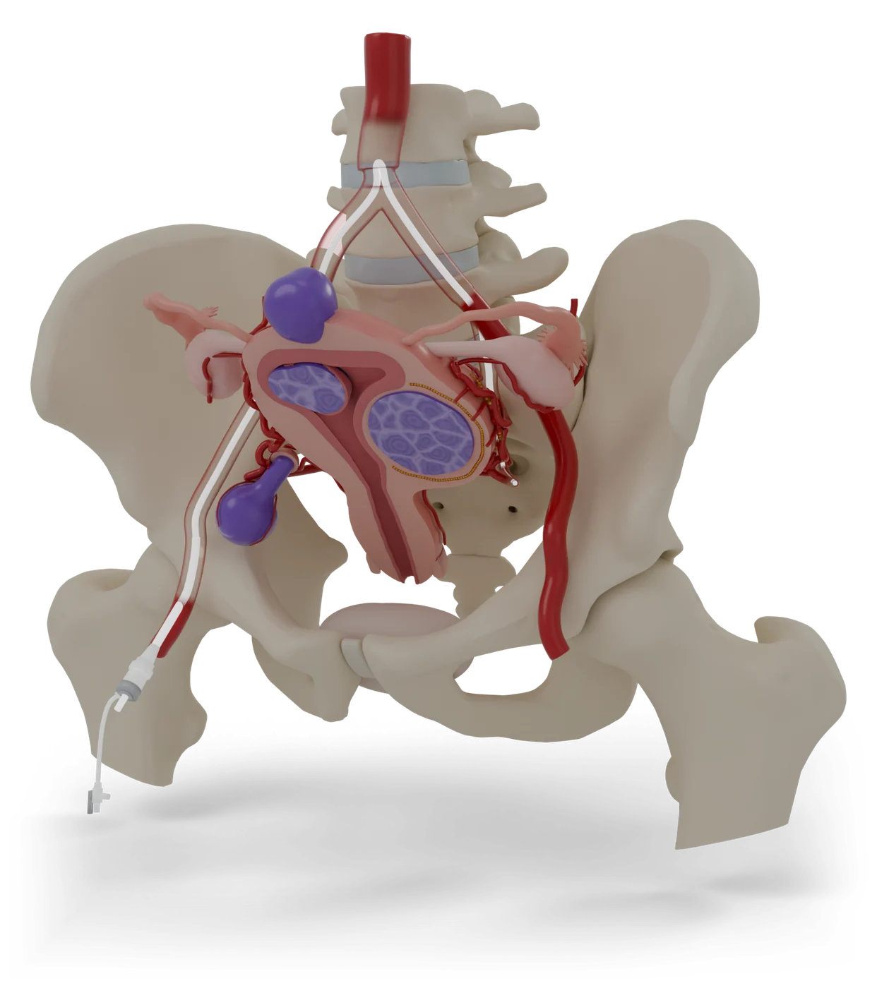
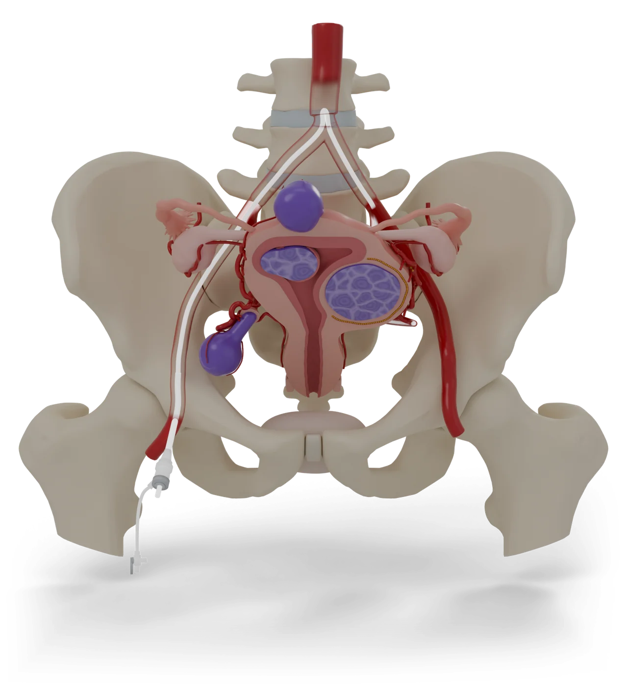
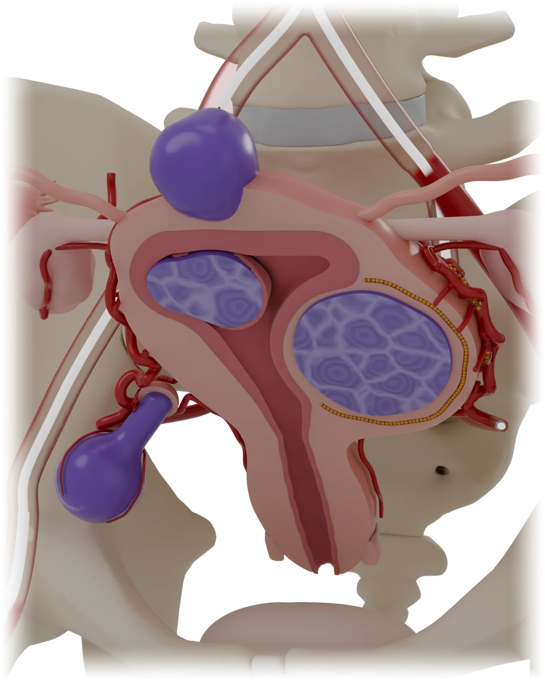
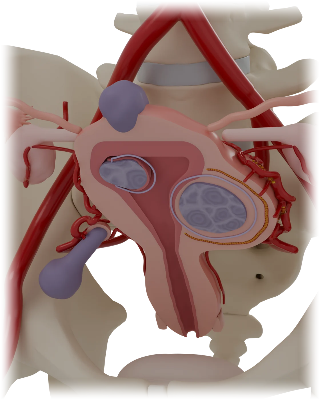
Colour key
- ArteriesAnatomy
- Uterine arteriesAnatomy
- FibroidsThe problem
- Fibroids, days afterAfter treatment
- Fibroids, 6 months laterAfter treatment
- Tubes and ovariesAnatomy
- DeviceWhat the doctor uses
- UterusAnatomy
- Uterus, front halfAnatomy
- Lining of the uterusAnatomy
- Top of the vaginaAnatomy
- Uterus, 6 months laterAfter treatment
- Lining, 6 months laterAfter treatment
- BladderAnatomy
- Fibroid size on the dayThe problem
- Particles, left sideWhat stays in
- Particles, right sideWhat stays in
Status
- 3D model
- Ready: 44 named parts, used by the patient guide.
- An earlier version was printed as a 23-part prototype. This model will be engineered for print next.
- Patient guide
- 12 steps in 6 parts, draft awaiting clinical review.
- Licence and credits
- Built from Z-Anatomy (CC BY-SA 4.0), based on BodyParts3D. Changed: simplified, recoloured, procedure parts added. Licence for this model: being confirmed before launch.
The story the model tells
- 1First, a quick look at the uterus and fibroidsWhat it treats
- 2Where a fibroid grows changes what it doesWhat it treats
- 3How UFE works: cut off the fibroids’ blood supplyHow it works
- 4Before the day: scans, tests and a planGetting ready
- 5On the day: relaxed, numbed and looked afterGetting ready
- 6A small opening at the groin or wristThe procedure
- 7A thin tube travels to the uterusThe procedure
- 8Tiny particles block the fibroids’ blood supplyThe procedure
- 9The tube comes out and the opening sealsThe procedure
- 10The first days: cramps are expectedAfterwards
- 11Six months later, the fibroids are smallerAfterwards
- 12Risks, explained plainlyRisks
Parts
| Part | Side | Shows |
|---|---|---|
| Aorta and iliac arteries | Anatomy | |
| Left uterine artery | Left | Anatomy |
| Right uterine artery | Right | Anatomy |
| Uterine artery branches | Anatomy | |
| Left ovarian artery | Left | Anatomy |
| Right ovarian artery | Right | Anatomy |
| Vessels feeding the fibroids | Anatomy | |
| Uterus | Anatomy | |
| Uterus, front half | Anatomy | |
| Lining of the uterus | Anatomy | |
| Top of the vagina | Anatomy | |
| Uterus, 6 months later | After treatment | |
| Lining, 6 months later | After treatment | |
| Left ovary | Left | Anatomy |
| Right ovary | Right | Anatomy |
| Left fallopian tube | Left | Anatomy |
| Right fallopian tube | Right | Anatomy |
| Bladder | Anatomy | |
| Fibroid size on the day | The problem | |
| Doorway tube (sheath) | What the doctor uses | |
| Catheter | What the doctor uses | |
| Particles, left side | Left | What stays in |
| Particles, right side | Right | What stays in |
| Fibroid in the wall | The problem | |
| Fibroid in the wall, days after | After treatment | |
| Fibroid in the wall, 6 months later | After treatment | |
| Fibroid bulging inside | The problem | |
| Fibroid bulging inside, days after | After treatment | |
| Fibroid bulging inside, 6 months later | After treatment | |
| Fibroid on the outside | The problem | |
| Fibroid on the outside, days after | After treatment | |
| Fibroid on the outside, 6 months later | After treatment | |
| Fibroid on a stalk | The problem | |
| Fibroid on a stalk, days after | After treatment | |
| Fibroid on a stalk, 6 months later | After treatment |