Nephrostomy tube
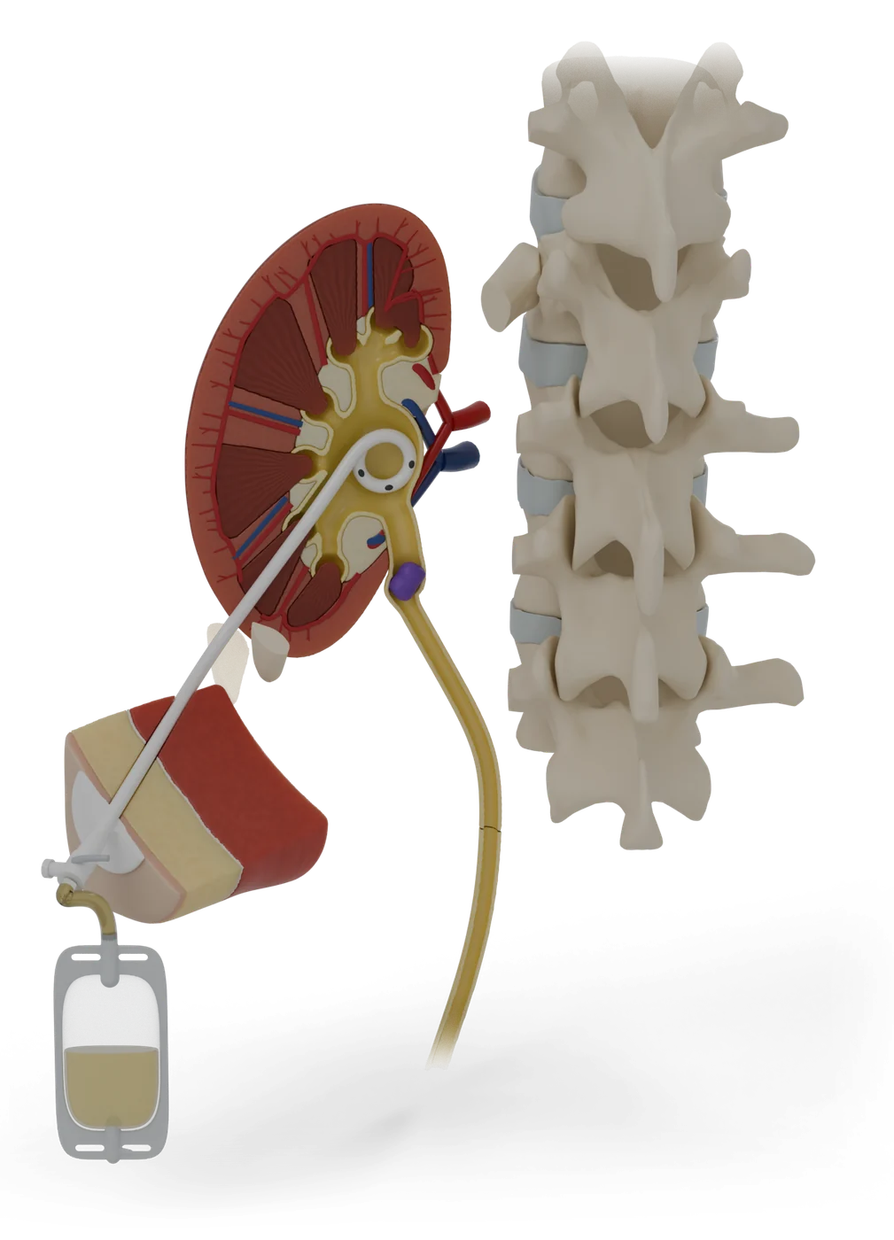
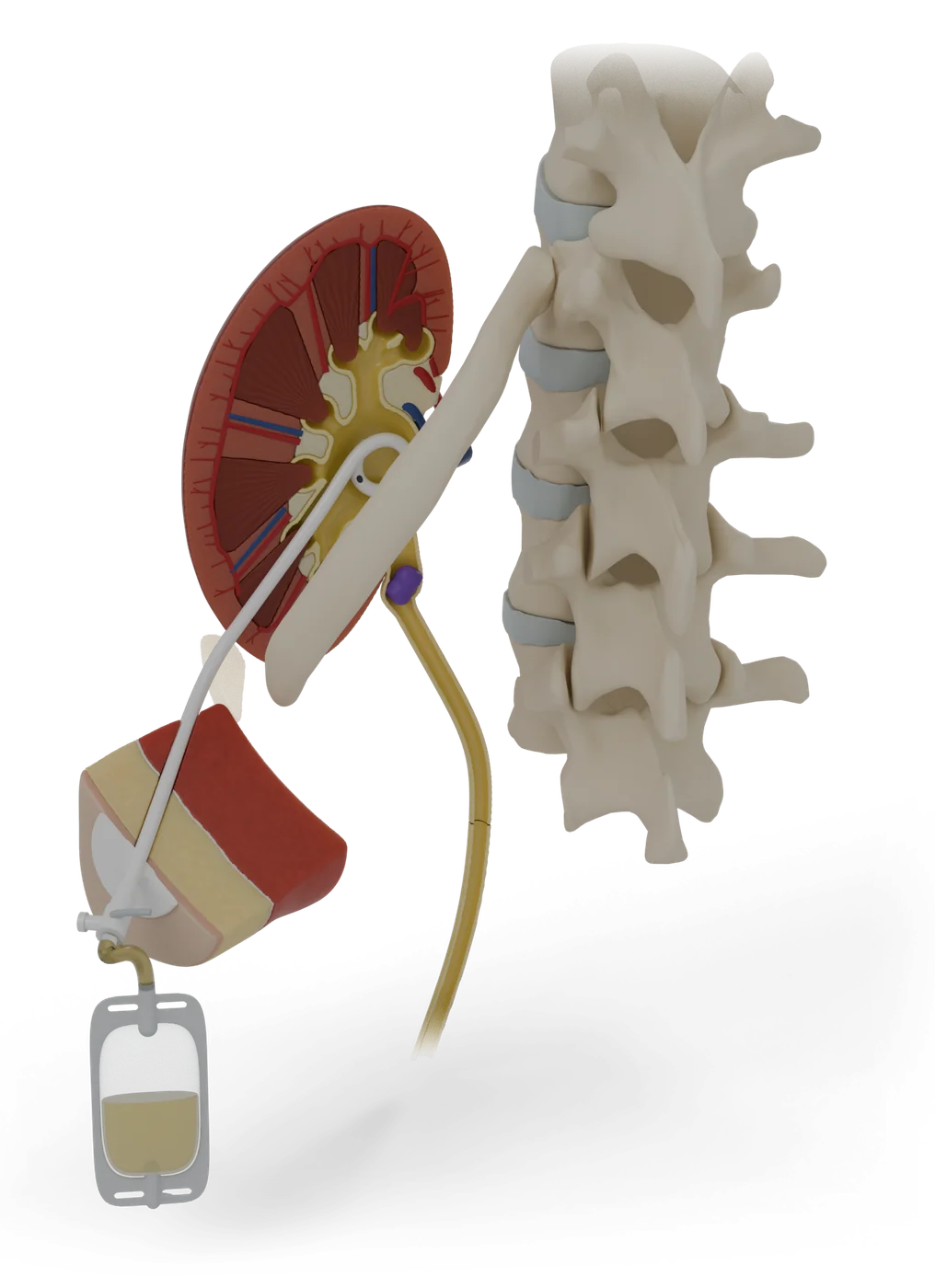
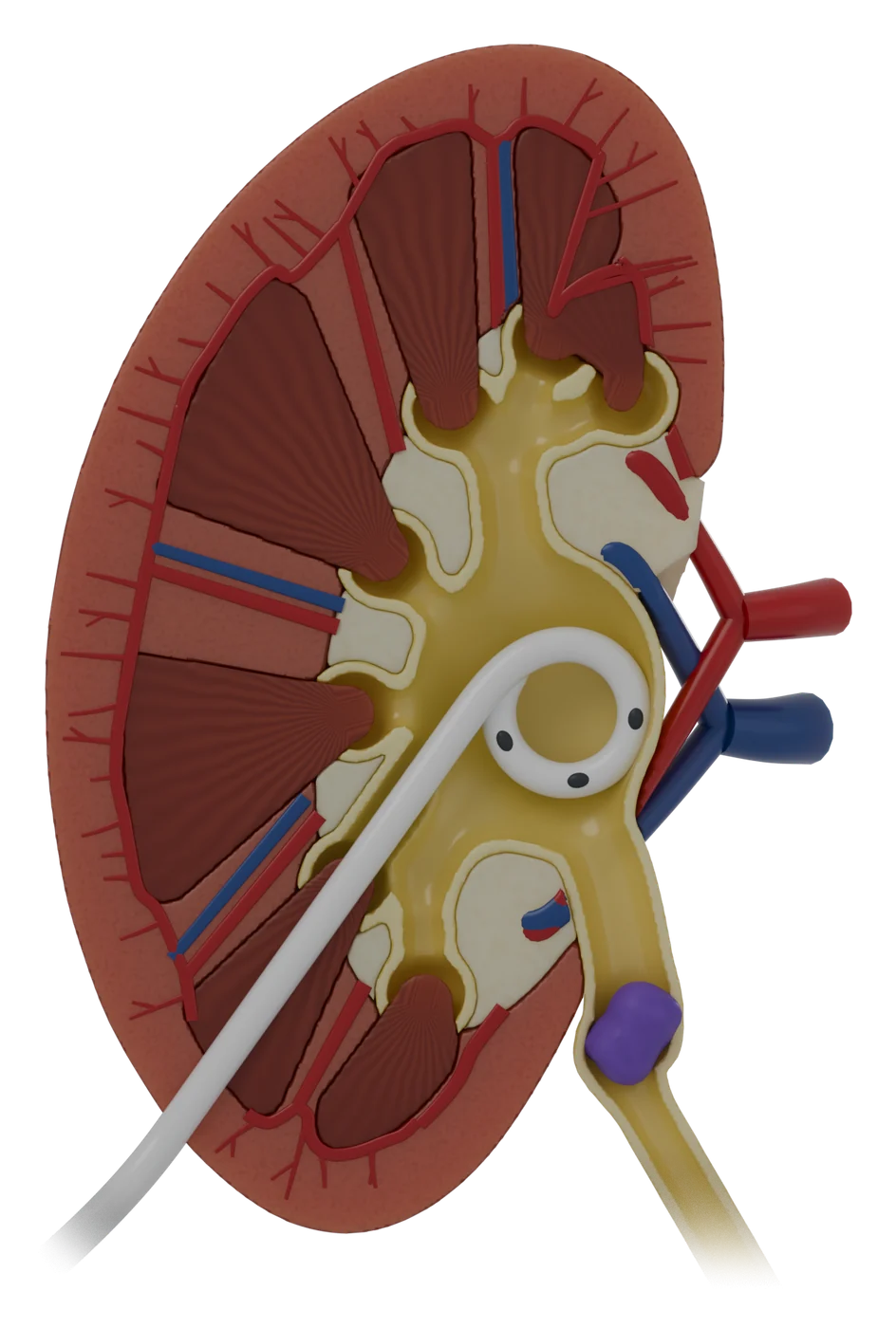
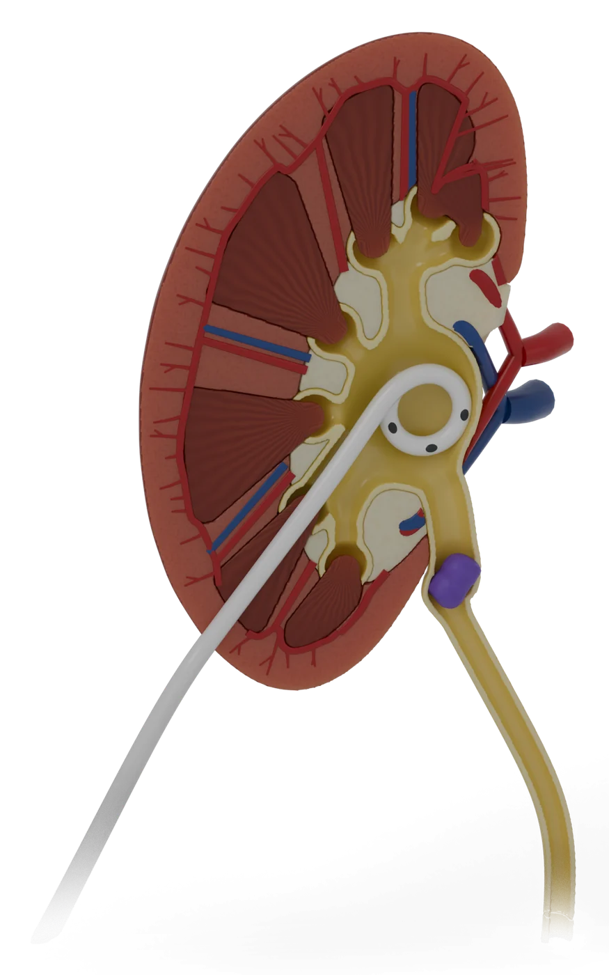
A thin tube through your back drains urine from a blocked kidney into a bag, to ease the pressure.
Colour key
- Blocked kidneyAnatomy
- Draining kidneyAnatomy
- Urine pathAnatomy
- Back half of the swollen kidneyAnatomy
- Back half of the kidneyAnatomy
- The blockageThe problem
- Planned entry pointWhat the doctor uses
- Nephrostomy tubeWhat the doctor uses
- Drainage tubing and tapWhat the doctor uses
- Fixation pad and dressingWhat the doctor uses
- Drainage bagWhat the doctor uses
- Urine in the bagAnatomy
- NeedleWhat the doctor uses
- GuidewireWhat the doctor uses
- Right kidneyAnatomy
- Right ureterAnatomy
- BladderAnatomy
Status
- 3D model
- Ready: 34 named parts, used by the patient guide.
- To come, from the same files as the on-screen model.
- Patient guide
- 12 steps in 5 parts, draft awaiting clinical review.
- Licence and credits
- Built from Z-Anatomy (CC BY-SA 4.0), based on BodyParts3D. Changed: simplified, recoloured, procedure parts added. Licence for this model: being confirmed before launch.
The story the model tells
- 1Your kidneys make urine all dayWhat it treats
- 2A blockage makes the kidney swellWhat it treats
- 3A tube gives the urine a way outHow it works
- 4What the model showsHow it works
- 5Getting readyThe procedure
- 6You lie face down, awake and numbThe procedure
- 7A thin needle goes into the kidneyThe procedure
- 8A wire guides the soft tube into placeThe procedure
- 9The tube is fixed to your skin and joined to a bagThe procedure
- 10The kidney can drain againAfterwards
- 11Living with the tube and bagAfterwards
- 12Risks, explained plainlyRisks
Parts
| Part | Side | Shows |
|---|---|---|
| Kidney, blocked and swollen (cut open) | Anatomy | |
| Swollen urine funnel and ureter | Anatomy | |
| Back half of the swollen kidney | Anatomy | |
| Kidney artery | Anatomy | |
| Kidney vein | Anatomy | |
| Kidney (cut open) | Anatomy | |
| Urine funnel and ureter | Anatomy | |
| Back half of the kidney | Anatomy | |
| Kidney artery | Anatomy | |
| Kidney vein | Anatomy | |
| The blockage | The problem | |
| Ureter (to the bladder) | Anatomy | |
| Planned entry point | What the doctor uses | |
| Nephrostomy tube | What the doctor uses | |
| Drainage tubing and tap | What the doctor uses | |
| Fixation pad and dressing | What the doctor uses | |
| Drainage bag | What the doctor uses | |
| Urine in the bag | Anatomy | |
| Needle | What the doctor uses | |
| Guidewire | What the doctor uses | |
| Right kidney | Right | Anatomy |
| Right ureter | Right | Anatomy |
| Bladder | Anatomy |