Endovascular aneurysm repair
Fixes a bulge in the body’s main artery from the inside, through small openings at both groins.
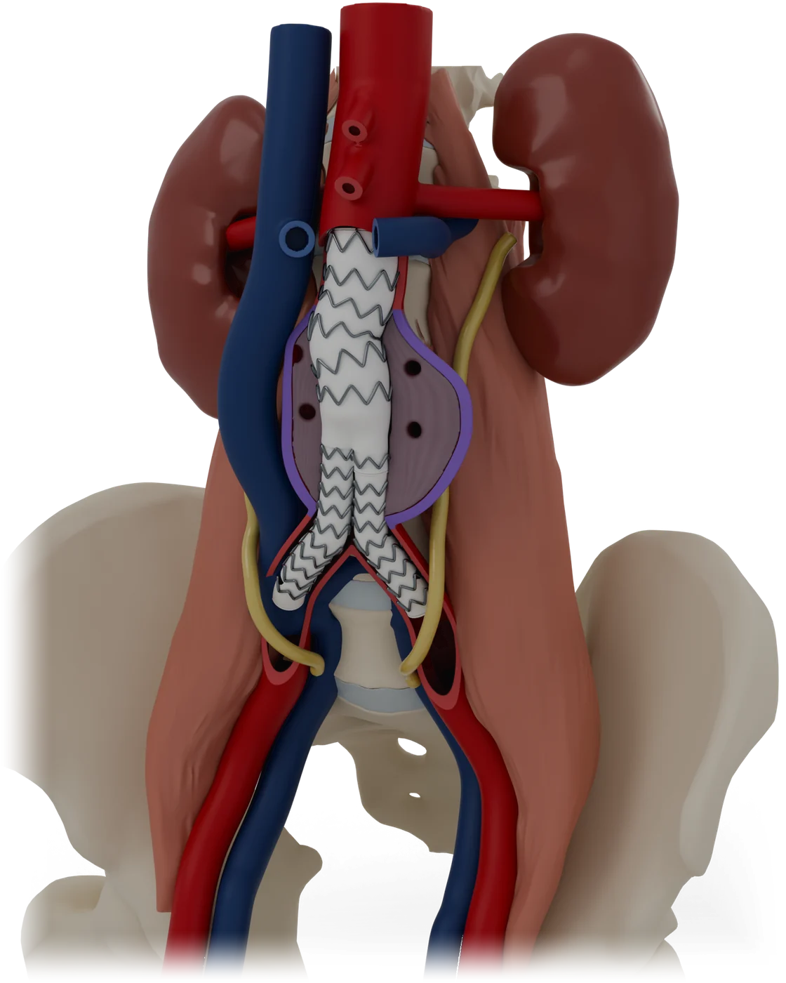
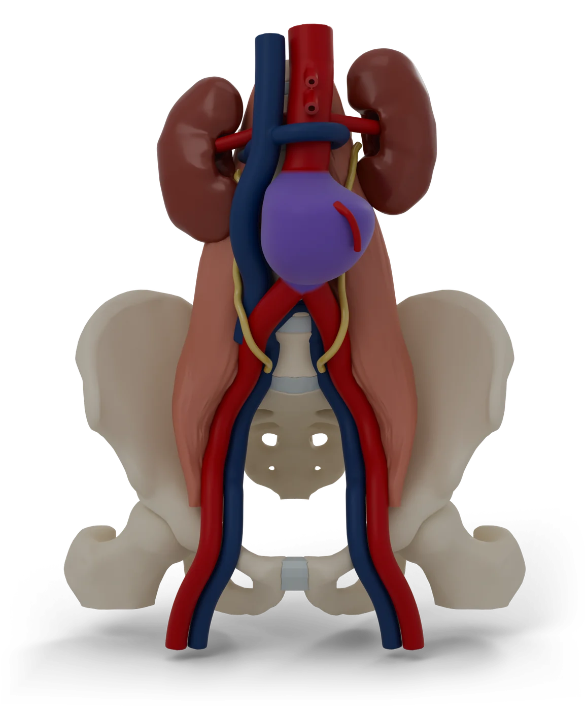
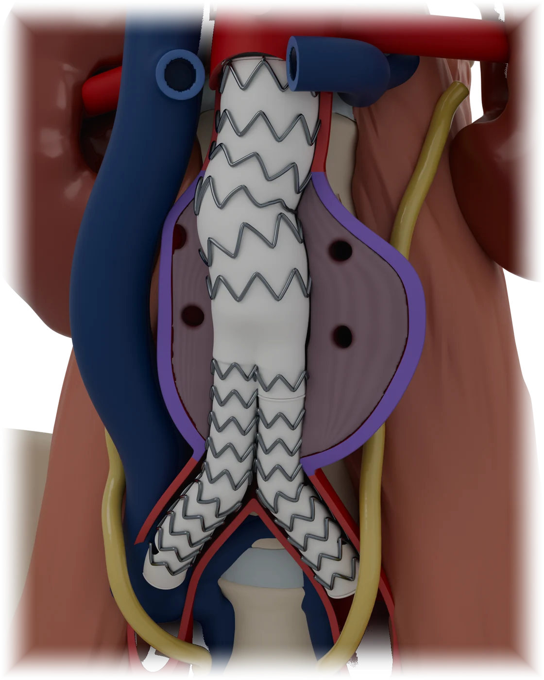
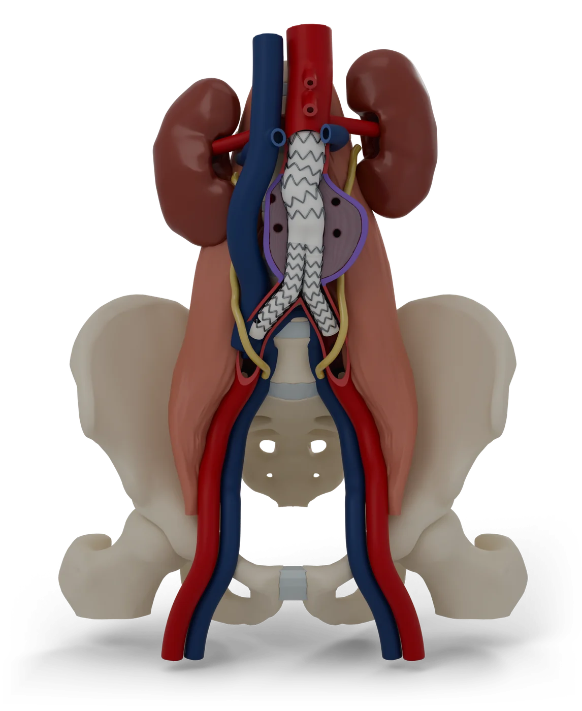
Colour key
- ArteriesAnatomy
- AneurysmThe problem
- Stent graftWhat stays in
- BonesAnatomy
- VeinsAnatomy
- SheathsWhat the doctor uses
- KidneysAnatomy
- Urine tubes (ureters)Anatomy
- Groin crease band (inguinal ligament)Anatomy
- Folded graft in its delivery tubeWhat the doctor uses
- Moulding balloonWhat the doctor uses
- Guide wireWhat the doctor uses
- Aneurysm, a year laterAfter treatment
- Scan slice, todayThe problem
- Scan slice, a year laterAfter treatment
Status
- 3D model
- Ready: 31 named parts, used by the patient guide.
- To come, from the same files as the on-screen model.
- Patient guide
- 12 steps in 5 parts, draft awaiting clinical review.
- Licence and credits
- Built from Z-Anatomy (CC BY-SA 4.0), based on BodyParts3D. Changed: simplified, recoloured, procedure parts added. Licence for this model: being confirmed before launch.
The story the model tells
- 1The aorta is your body’s main arteryWhat it treats
- 2An aneurysm is a weak spot that bulgesWhat it treats
- 3A large bulge can burst, so it is fixed firstWhat it treats
- 4A stent graft lines the artery from the insideHow it works
- 5The graft comes in pieces that join inside youHow it works
- 6Getting ready: scans, tests and planningThe procedure
- 7The way in is through both groinsThe procedure
- 8The main body opens just below the kidney arteriesThe procedure
- 9The second leg joins, and the seal is checkedThe procedure
- 10The tubes come out and the groins are closedThe procedure
- 11The bulge is checked for the rest of your lifeAfterwards
- 12Risks, explained plainlyRisks
Parts
| Part | Side | Shows |
|---|---|---|
| Lower spine | Anatomy | |
| Spinal discs | Anatomy | |
| Hip bones | Anatomy | |
| Tops of the thigh bones | Anatomy | |
| Kidneys | Anatomy | |
| Urine tubes (ureters) | Anatomy | |
| Aorta | Anatomy | |
| Kidney arteries | Anatomy | |
| Aneurysm | The problem | |
| Aneurysm, front wall | The problem | |
| Old clot inside the bulge | The problem | |
| Artery front wall | Anatomy | |
| Leg arteries (iliac and femoral) | Anatomy | |
| Groin artery front wall | Anatomy | |
| Small side artery | Anatomy | |
| Main vein and leg veins | Anatomy | |
| Left kidney vein | Left | Anatomy |
| Groin crease band (inguinal ligament) | Anatomy | |
| Stent graft, main body | What stays in | |
| Stent graft, second leg | What stays in | |
| Folded graft in its delivery tube | What the doctor uses | |
| Moulding balloon | What the doctor uses | |
| Sheath, right groin | Right | What the doctor uses |
| Sheath, left groin | Left | What the doctor uses |
| Guide wire | Right | What the doctor uses |
| Guide wire | Left | What the doctor uses |
| Aneurysm, a year later | After treatment | |
| Scan slice, today | The problem | |
| Scan slice, a year later | After treatment |